神经鞘黏液瘤
Nerve Sheath Myxoma
刘正智
发布时间:2016-08-03 22:27:01
同义词(或曾用名):真皮神经鞘黏液瘤
概述:
发病部位:远端肢体,手和足部最常见
诊断要点:
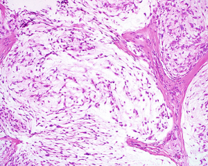
2. 低倍镜下呈大小不等的小叶结构,小叶间为纤维结缔组织间隔;
3. 小叶主要由星状或梭形细胞组成,偶见圆形上皮样细胞;
4. 瘤细胞排列疏松,间质内含有大量的透明质酸或硫酸黏液,AB染色为阳性;
5. 瘤细胞的胞质呈淡嗜伊红色,常见细长的胞质突起,在小叶或结节的边缘肿瘤细胞常见胞浆内空泡形成印戒样排列;
6. 小叶内的瘤细胞无异型性,核分裂像罕见。
免疫组织化学染色:
鉴别诊断:
1. 细胞性neurothekeoma(NTK):多见于头颈部和上肢,罕见于手和足;肿瘤边界不清楚,呈微结节装生长,瘤细胞上皮样细胞为主伴有丰富的浅染嗜酸性胞浆,黏液变性通常不广泛,免疫组化染色不表达S100蛋白和SOX10,表达CD68和CD10,MITF等;
2. 丛状神经纤维瘤:通常为NF1患者,丛状结构周围无厚的纤维性包膜围绕,肿瘤罕见广泛的黏液变性,免疫组化除了表达S100之外,尚可见CD34阳性的纤维母细胞和NF阳性的轴突。
预后:
治疗:
参考文献:
1.Suarez A et al: Immunohistochemical analysis of KBA.62 in 18 neurothekeomas: a potential marker for differentiating neurothekeoma, but a marker that may lead to confusion with melanocytic tumors. J Cutan Pathol. 41(1):36-41, 2014
2.Stratton J et al: Cellular neurothekeoma: analysis of 37 cases emphasizing atypical histologic features. Mod Pathol. Epub ahead of print, 2013
3.Sheth S et al: Differential gene expression profiles of neurothekeomas and nerve sheath myxomas by microarray analysis. Mod Pathol. 24(3):343-54, 2011
4.Vered M et al: Classic neurothekeoma (nerve sheath myxoma) and cellular neurothekeoma of the oral mucosa: immunohistochemical profiles. J Oral Pathol Med. 40(2):174-80, 2011
5.Nishioka M et al: Nerve sheath myxoma (neurothekeoma) arising in the oral cavity: histological and immunohistochemical features of 3 cases. Oral Surg Oral Med Oral Pathol Oral Radiol Endod. 107(5):e28-33, 2009
6.Fetsch JF et al: Neurothekeoma: an analysis of 178 tumors with detailed immunohistochemical data and long-term patient follow-up information. Am J Surg Pathol. 31(7):1103-14, 2007
7.Reimann JD et al: Myxoid dermatofibrosarcoma protuberans: a rare variant analyzed in a series of 23 cases. Am J Surg Pathol. 31(9):1371-7, 2007


