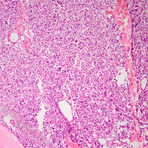
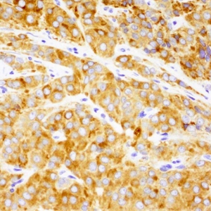
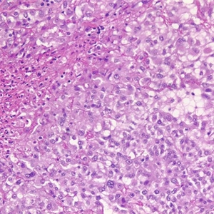
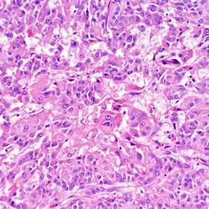
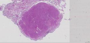
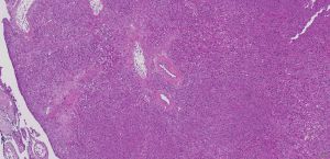
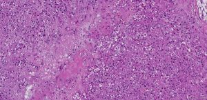
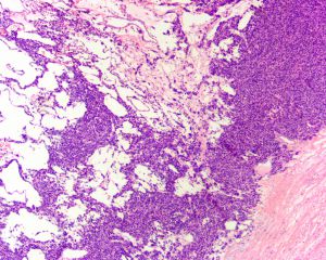
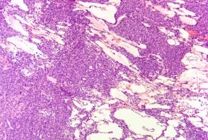
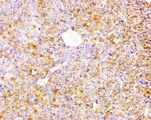
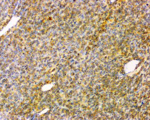
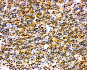
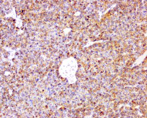
2.低倍镜下肿瘤界限清楚,包膜完整,最常表现为弥漫性生长,部分病例由胶原纤维分割成大的结节,少见病例可见岛状、梁状、假管状、带状梭形细胞样或微囊状结构;
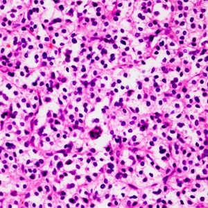
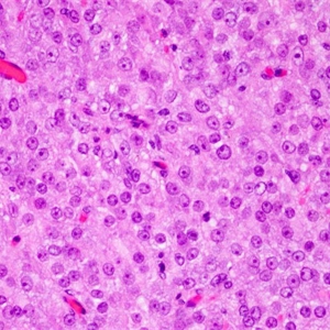
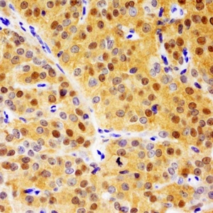
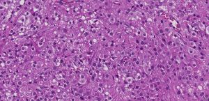
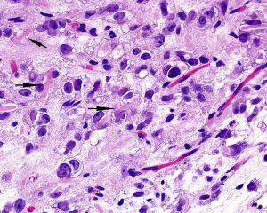
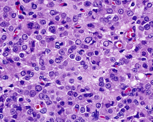
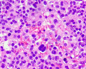
3.肿瘤细胞通常呈形态一致的圆形,具强嗜酸性胞浆,30%病例胞浆内可见棒状的Reink结晶,15%病例可见脂褐素;有些病例可见多少不等的泡沫状胞浆;
4.细胞核通常圆形,核仁明显,位于细胞核中央,局部核染色质空泡状呈毛玻璃样核特征,细胞异型轻微或无异型,核分裂像少见;
5.偶尔可见灶状脂肪化生、钙化及骨化;
6.间质有丰富的血管网结构,亦可透明变性,偶尔间质水肿,可见砂粒体形成;
7.约5%的表现出恶性组织学特征,存在以下2个或以上特征提示恶性:肿瘤直径>5cm, 浸润性生长,弥漫的细胞非典型性,核分裂象>3个/10HPF, 血管浸润和/或存在肿瘤性坏死。
2.间质细胞增生:通常为镜下表现,直径<0.5cm,罕见表现为占位性病变,但通常为多灶性增生;
3.成人颗粒细胞瘤:Melan-A通常阴性;
4. 软斑病:累及生精小管和间质细胞,黄色肉芽肿性间质反应,病变细胞内可见特征性的Michaelis-Gutmann小体;
5. 卵黄囊瘤:瘤细胞核较幼稚,核分裂象多,免疫组化表达SALL4。
Suardi N, Strada E, Colombo R, et al. Leydig cell tumour of the testis: presentation, therapy, longterm follow-up and the role of organ-sparing surgery in a single-institution experience. BJU Int 2009;103(2):197–200.
Ulbright TM, Srigley JR, Hatzianastassiou DK, et al. Leydig cell tumors of the testis with unusual features: adipose differentiation, calcification with ossification, and spindle-shaped tumor cells. Am J Surg Pathol 2002;26(11):1424–33.
Kim I, Young RH, Scully RE. Leydig cell tumors of the testis. A clinicopathological analysis of 40 cases and review of the literature. Am J Surg Pathol 1985;9(3):177–92.
