梭形细胞脂肪瘤
Spindle cell lipoma
刘正智 夏成青
发布时间:2016-10-08 07:09:31
概述:
发病部位:境界清楚的皮下结节,常位于老年男性颈部和背部。
诊断要点:
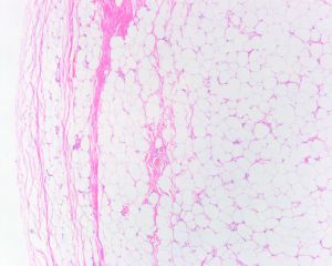
1.境界清楚的皮下结节,无痛性,生长缓慢,常位于老年男性颈部和背部;肿块通常3-5cm,很少>10cm;
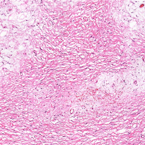
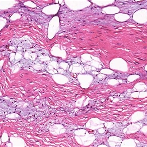
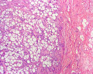
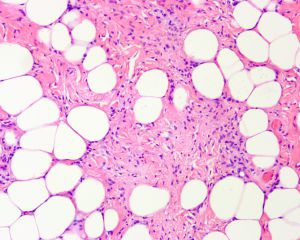
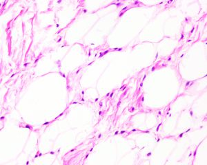
2.在脂肪瘤的背景上可见小的、核深染的梭形和圆形细胞及细胞核放射状排列呈“花环状”的多核巨细胞;
3.可能是家族性;
4.细胞遗传学:16q13-qter缺失,13q12和13q14-22缺失。
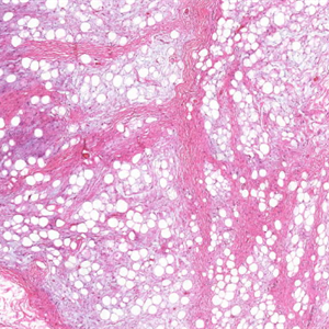
5.粘液样基质是常见的,可能是广泛的。
6.肥大细胞非常常见,几乎是恒定的。
7.厚壁血管可显示血管周围透明质变性或纤维化或在局灶性鹿角状增生中生长。
8.多形性脂肪瘤:
以多核巨细胞为特征,嗜酸性胞质致密,外周排列细胞核(小花细胞);
纺锤状细胞、绳索样胶原、肥大细胞和脂肪组织通常也存在;
可能很少含有多空泡化脂肪细胞;
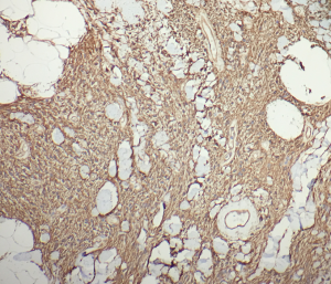
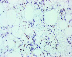
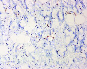
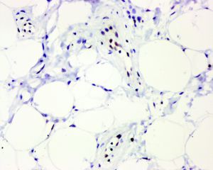
免疫组织化学染色:
分子标记:
鉴别诊断:
1.非典型性脂肪瘤性肿瘤/高分化脂肪肉瘤:位于肢体或腹膜后深部软组织,肿瘤内可见核深染的异型间质细胞,肿瘤组织内缺乏绳索样胶原纤维结构-是鉴别的重要依据,FISH常有MDM2、CDK4基因扩增;
2.结节性筋膜炎:常见中青年,好发于前臂屈侧皮下或更深部,病程多在1-2周,黏液间质内见星状或梭形细胞,SMA(+),CD34(-);
3.血管肌纤维母细胞瘤:好发于中青年女性外阴,有交替分布的细胞丰富区和细胞稀疏区组成,肿瘤内含大量扩张的小到中等大薄壁血管,Vimentin、desmin、ER、PR(+);
4. 非典型梭形细胞脂肪瘤样肿瘤:与梭形细胞脂肪瘤具有重叠的组织学,免疫表达以及遗传学特征,但 常见浸润性生长,其内可见脂肪母细胞。
治疗:
参考文献:
1.Cheah A et al: Spindle cell/pleomorphic lipomas of the face: an under-recognized diagnosis. Histopathology. 66(3):430-7, 2015
2.Creytens D et al: Atypical spindle cell lipoma: a clinicopathologic, immunohistochemical, and molecular study emphasizing its relationship to classical spindle cell lipoma. Virchows Arch. ePub, 2014
3.Lau SK et al: Spindle cell lipoma of the tongue: a clinicopathologic study of 8 cases and review of the literature. Head Neck Pathol. ePub, 2014
4.Chen BJ et al: Loss of retinoblastoma protein expression in spindle cell/pleomorphic lipomas and cytogenetically related tumors: an immunohistochemical study with diagnostic implications. Am J Surg Pathol. 36(8):1119-28, 2012
5.Flucke U et al: Cellular angiofibroma: analysis of 25 cases emphasizing its relationship to spindle cell lipoma and mammary-type myofibroblastoma. Mod Pathol. 24(1):82-9, 2011
6.Wood L et al: Cutaneous CD34+ spindle cell neoplasms: Histopathologic features distinguish spindle cell lipoma, solitary fibrous tumor, and dermatofibrosarcoma protuberans. Am J Dermatopathol. 32(8):764-8, 2010
7.Sachdeva MP et al: Low-fat and fat-free pleomorphic lipomas: a diagnostic challenge. Am J Dermatopathol. 31(5):423-6, 2009
